Your heart is a muscle about the size of a fist, located a little left of the middle of your chest. Your heart works like a pump, sending oxygen-rich blood around your body.
The Structure of a Heart
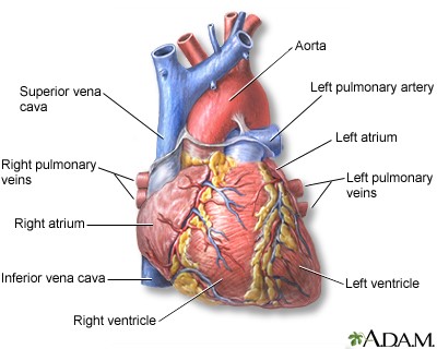
The structures of your heart include the ventricles, atria, arteries and veins. Arteries carry blood away from your heart while veins carry blood back to your heart.
Your heart is composed of 2 independent pumping systems - one on the right side, and the other on the left. Each has 2 chambers, an atrium (upper chamber) and a ventricle (lower chamber). The ventricles are the major pumps in your heart.
The Right Side of Your Heart
The right system receives blood from the veins of the whole body. This is "used" blood which is poor in oxygen and rich in carbon dioxide.
- The right atrium is the first chamber that receives blood. The chamber expands as its muscles relax to fill with blood that has returned from the body.
- The blood enters a second, more muscular, chamber called the right ventricle. The right ventricle is one of your heart's 2 major pumps. Its function is to pump the blood into your lungs.
- Your lungs restore oxygen to the blood and exchange it with carbon dioxide, which you exhale.
The Left Side of Your Heart
The left system receives blood that is now rich in oxygen from your lungs.
- The oxygen-rich blood returns through veins coming from your lungs (pulmonary veins) to your heart.
- Your heart receives the oxygen-rich blood from your lungs in the left atrium, the first chamber on the left side.
- The blood then moves to the left ventricle, a powerful muscular chamber that pumps the blood back out to your body. The left ventricle is the strongest of your heart's pumps. Its thicker muscles need to perform contractions powerful enough to force the blood to all parts of your body.
The Heart Valves
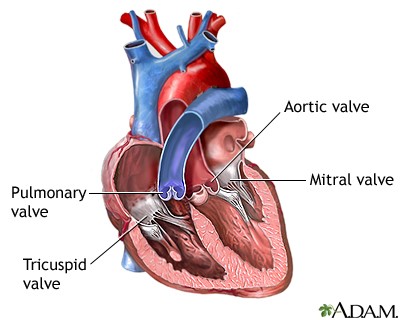
Valves are muscular flaps that open and close so that blood will flow in the right direction. There are 4 valves in your heart:
- The tricuspid valve regulates blood flow between the right atrium and the right ventricle.
- The pulmonary valve allows blood to flow from the right ventricle to the lungs.
- The mitral valve regulates blood flow between the left atrium and the left ventricle.
- The aortic valve allows blood to flow from the left ventricle to the aorta – the major artery that feeds blood to the entire body.
These valves open up just enough so that blood can flow through. Then they close, keeping blood from flowing backward. Heart valve disease happens when your heart valves don’t work the way they should because of damage or a defect.
Your Heart's Electrical System
Your heart has an electrical system that makes sure it contracts (squeezes) in an orderly way.
The electrical impulse that signals your heart to contract begins in an area of the heart called the sinoatrial node (also called the sinus node or SA node). This is your heart’s natural pacemaker.
The signal leaves the SA node and travels through the heart along a set electrical pathway. Different nerve messages signal your heart to beat slower or faster.
Arrhythmias are caused by problems with the heart’s electrical conduction system.
Heart and Vascular Center
Wake Forest Baptist’s Heart and Vascular Center combines cardiology, cardiothoracic surgery and vascular surgery to provide a multidisciplinary team approach to patient- and family-centered care. At the Heart and Vascular Center, our philosophy is clear: patients come first. We offer the latest in technology, devices and medication combined with personalized care, to offer life-changing vascular and heart disease treatments.